


A team of researchers from the Duke-NUS Medical School and Singapore General Hospital recently repurposed a non-invasive cancer detection method in order to track dengue infection in mouse models in real time.
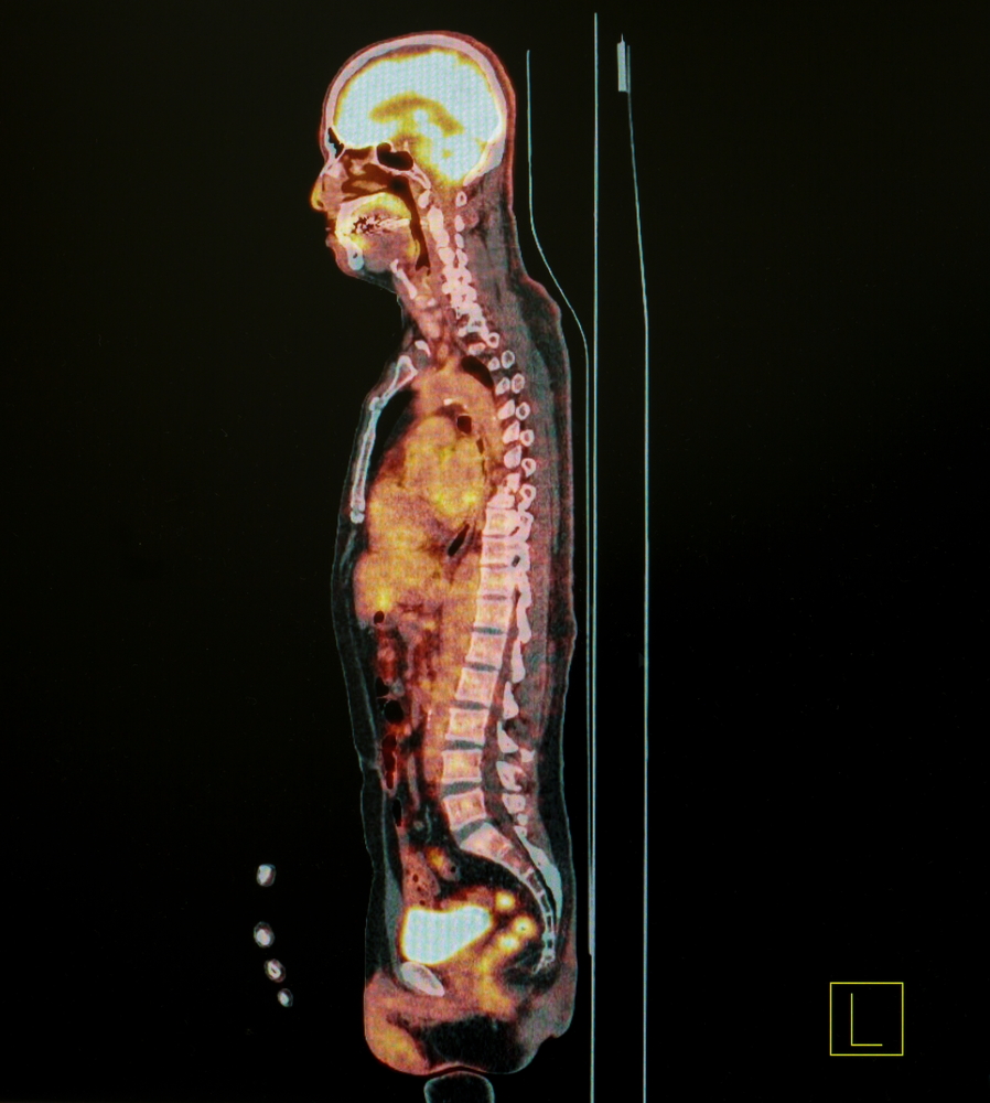
Typically used to detect solid tumors, positron emission tomography (PET) has the ability to track where glucose is being taken up by cells in a process similar to that of radar. Fluorodeoxygluscose (FDG), a radioactive version of glucose, can be seen using PET technology when injected directly into mice and absorbed by their cells.
Since inflammation of the small and large intestines has been known to occur in mice when infected with dengue, along with a cellular uptake of glucose and FDG increases, the research teams utilized PET-FDG to visualize inflammation as a marker of dengue infection.
“To our knowledge, this is the very first time PET has been systematically evaluated in the field of acute viral infectious disease,” Ann-Marie Chacko, a Duke-NUS assistant professor and lead author of the study, said.
The PET-FDG method allowed researchers to observe inflammation in the spleen, small intestine, and large intestine of dengue-infected mice, which subsided after antivirals were administered. By tracking glucose uptake, the method helped researchers predict the progression and severity of infection, along with the effectiveness of the treatment.
“Traditionally, in research, the amount of virus in the blood is measured and used as an indicator of disease severity,” Jenny Low, a senior consultant with Singapore General Hospital, said. “What makes the findings of this study so groundbreaking is that we may have a non-invasive way to track dengue infections in our patients more accurately during clinical trials to better measure if the experimental treatment given is effective.”
The study was published in a recent issue of the Journal of Clinical Investigation Insight.